Dr. Renat
Letfullin
e-mail:
Letfullin@rose-hulman.edu
My research program is highly interdisciplinary and combines techniques from the fields of biophysics, nanomedicine, nanoscience, wave optics, laser physics, and aerosol physics, including optics of nanoparticles. Selected projects are mentioned below.
My research in the Biophysics area includes:
� A wave electrodynamics model of laser light interaction with nanosized biological scattering centers. This model describes the propagation of the wave into strongly scattering biological tissue based on the solution of the Maxwell wave equations. The primary scattering centers are thought to be the collagen fiber network of the extracellular matrix, the mitochondria, and other intracellular substructures, all with dimensions smaller than optical wavelengths. Another source of scattering centers is the artificially injected nanoparticles into the biotissue and the cells: liposomes (30-90 nm), gold nanoparticles (2-250 nm), neutral red-stained particles (30-500 nm), and polystyrene nanoparticles (30-80 nm).
� Exact diffraction simulations of the integral and differential optical properties of nanosized scattering centers below the diffraction limit. The amplitude-phase delay of an electromagnetic wave due to (1) scattering on the nanocenters described above and (2) the photo-thermal-induced inhomogeneities of the medium, is calculated on the basis of the wave equation and Mie diffraction theory. I conduct exact diffraction calculations of the integral and differential nanoparticle optical properties (such as scattering, absorption, attenuation cross sections, and scattering amplitude functions) below the diffraction limit for a broad range of the Mie parameter and temperature intervals.
� Non-destructive optical techniques for diagnostics of the biological/particle nanostructures. One technique is two-color laser transmissometry. Another technique for phase control of the nanoparticles characteristics is based on the interference strips displacement method, which can be realized with asymmetric Mach-Zehnder interferometer.
� Theoretical model for laser heating and evaporation of nanostructures and biological tissue. This model takes into account the temperature dependences of the optical and thermo-physical parameters of the particles and surrounding medium, as well as the real-time shape of the laser pulse. These calculations can be performed at different heat-mass transfer modes, such as the free molecular and diffusion modes.
� Laser Ablation and Optical Damage of the Cancer Cells by Nanoparticles. The characteristics of laser-induced cell damage depend on the rates of the basic processes taking place in the heated biological system containing the nanoparticles. These include the rates of the tissue inflammation � the first damage stage, the rates of the cancerous cells necrosis � the second damage stage, and the rates of evaporation and carbonizing of the cancer cells � the third damage stage. I develop an effective kinetic model of cancer-cell killing by laser-activated nanoheaters, which will include the most important physical-chemical kinetic and biological features. This model will predict the damage depth and the exposure time for each stage of laser/nanoparticle damage.
My research in the Nanomedicine area includes development of the Theory and Simulation Techniques for Selective Cancer Nanotherapy:
� Surface plasmon resonance as a cancer biomarker detection technology. Surface plasmon resonance (SPR) has been investigated both for its fundamental importance and as a possible detection technique with biochemical applications. Recently, commercial biosensors have become available that rely on SPR for detecting various proteins and other DNA constituents. Together with undergraduate students I perform simulations of the integral and differential optical properties (such as scattering, absorption and attenuation cross sections and scattering amplitude functions) of nanoparticles below the diffraction limit for a broad range of the wavelengths of transverse electromagnetic waves.
� Ultrashort laser pulse heating of nanostructures in cancer cells. Two models will be studied by the PI/co-PI to describe ultrashort laser pulse interactions with metal nanoparticles in time. The first is a two-temperature model which involves fast thermalization within the electron subsystem, non-uniform energy transfer to the lattice, and electron energy losses due to electron heat transport into the particle volume. The second is a one-temperature model based on a heat transfer equation written within a uniform heating approximation throughout the particle volume. Along with and participating students I perform the comparative simulations of temperature-time dynamics using these models for laser heating of metallic nanotargets in the femtosecond, picosecond and nanosecond regimes, thus providing an effective modeling method to explore the effects playing the determining role in the laser-matter interactions.
� Laser-induced explosion of nanoparticles. The strongly-enhanced SPR of noble metal nanoparticles at optical frequencies makes them excellent scatters and absorbers of visible and near-IR light. When the strongly-absorbing nanoparticles are irradiated by ultrashort laser pulses, their temperature rapidly reaches the thresholds for nonlinear effects and can cause an explosion of nanoparticles. This involves the generation of a shock wave of dense vapors expanding in the cell volume with supersonic velocity, producing sound waves and optical plasma. A phenomenological picture of these complex physical effects is shown schematically in Fig. 1.
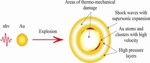
Figure 1. Laser-induced thermal explosion of a nanoparticle.
There are two main physical mechanisms that could lead to the laser-induced explosion of nanoparticles - (a) thermal explosion mode through electron-phonon excitation-relaxation and (b) Coulomb explosion mode through multiphoton ionization. I simulate the threshold laser energy density required for the realization of the thermal explosion of nanoparticles.
New Dynamic Modes in Selective Cancer Nanomedicine:
� Microbubble overlapping mode. Bioconjugated nanoparticles are selectively attached to chosen cellular targets, in particular to membrane receptors activated by other antibodies as shown in Fig. 2. When nanoparticles are irradiated by short laser pulses, they absorb the laser radiation and their temperature rises very quickly, reaching up to the threshold of microbubble formation in the surrounding liquid medium.
I�m studying a new dynamic mode for selective cancer treatment that involves the overlapping of bubbles inside the cell volume (shown in bottom part of Fig. 2). The microbubble overlapping mode around intracellular structures induced by short laser pulses can dramatically increase the efficiency of the cancer treatment as a result of the larger damage range and higher expenditure rate in comparison to a thermal damage mode.
� Nanoclusters aggregation mode. I propose a new mechanism for selective laser killing of abnormal cells by nanoclusters aggregated in the cell volume, as shown in Fig. 3(a). A cluster is a group of closely-located nanoparticles separated by the thickness of antibodies (10-30 nm), which has a typical size of 200-400 nm. Here, the effective therapeutic effect for cancer cell killing is achieved due to a large damage area at the relatively-low energy density of the incident laser pulse.
Figure 2. Principle of selective nanophotothermolysis in the laser-induced microbubble overlapping mode around gold nanoparticles.
� Thermal explosion mode � �nanobombs.� I propose yet another new mechanism for selective laser killing of abnormal cells by laser thermal explosion of single nanoparticles � nanobombs � delivered to the cells, as shown in Fig. 3(b). Thermal explosion of nanoparticles is realized when heat is generated within the strongly-absorbing target more rapidly than the heat can diffuse away. Here, the effective therapeutic effect for cancer cell killing is achieved due to nonlinear phenomena, which accompany the thermal explosion of the nanoparticles: generation of a strong shock wave with supersonic expansion of a dense vapor in the cell volume, producing sound waves and optical plasma.
Figure 3. Principle of the nanocluster aggregation mode (a) and thermal explosion mode (b) in selective nanophotothermolysis.
In the field of OPTICS:
� A New Optical Effect of Diffractive Multifocal Focusing of Radiation on a Bicomponent Diffraction System. This new optical effect can be observed in the near field (Fresnel zone), when the electromagnetic wave diffracts on the system of pinholes. The DMRF effect can dramatically increase the on-axis intensity of the diffracted wave without using traditional refraction elements, such as lenses, prisms, etc. This effect is occurs for a wide variety of radiation wavelengths and it does not disappear even in strong scattering media. This new phenomenon presents itself independent of ones interests in Optics, and it has great applied values in lasers.
� A New Type of Auto-Wave - Auto-Wave Effect of a Photon-Branched Chain Reaction. The investigation of photon-branched chain reactions (PBCR) may result in the discovery of a fundamentally new physical process, in which the photon plays the role of a direct participant in the chemical process, together with the atoms and the molecules. A PBCR is accompanied by directional energy evolution and under some conditions such a reaction can be capable of self-propagation along a certain direction. Essentially, a new auto-wave (self-wave) appears and energy is transfered in this wave by photons, which are simultaneously participants of the chemical process and its products. This auto-wave regime makes it possible to achieve a much stronger amplification of the energy of the initiating pulse.
� Effect of Giant Amplification of Wave Energy in a Two-Phase Active Medium. An optical effect of diffractive focusing of the initiating wave on an input aperture and auto-wave ignition of a photon-branched chain reaction can lead to a new effect of giant laser energy gain up to ~ 1011 in a two-phase active medium. This phenomenon has a threshold character and plays an important role in laser physics.
� Single Wave Scattering on Dispersed Particles. An amplitude-phase delay of an electromagnetic wave due to scattering on dispersed particles and on photo-thermal induced inhomogeneities of the medium are being calculated on the basis of the wave equation and Mie diffraction theory. These studies are important for exact calculations of optical properties of particles (the scattering, absorption and attenuation cross-sections in a broad range of the Mie parameter, that is for different sizes and materials of particles, and for a wide variety of radiation wavelengths) and for modeling of wave propagation in scattering and absorbing media.
In the field of LASER PHYSICS:
� Physical Conceptions for Developing High-Power Pulsed Chemical Lasers Based on a Photon-Branched Chain Reaction. The main goals of this study are to develop the principles for creating small scale lasers based on a photon-branched chain reaction and to obtaining the key data for construction of high-power pulsed chemical lasers, which do not consume the energy of external sources. The design of self-contained compact portable laser systems with output energy in a pulse of 2 kJ is necessary for many practical civil applications.
� Cavity Optics. The present study is devoted to an analysis of an auto-wave photon-branched process in an unstable telescopic cavity and to the dynamics of cavity field propagation in a two-phase active medium, based on a simultaneous solution of the equations of laser chemical kinetics and the wave equation.
� Compact Self-Contained Pulsed Laser Based on an Auto-Wave Photon-Branched Chain Reaction. Predicted effects of auto-wave spreading of PBCR and giant laser energy gain allow us to get high amplification of energy in the relatively small volumes of an active medium. In this study I propose and design a self-contained compact pulsed HF laser, with small linear sizes for the unstable telescopic cavity, which can be initiated by a small microjoule master oscillator powered by an accumulator.
� New Principles for an Effective Optical Initiation of a Pulsed Laser Based on an Auto-Wave PBCR. At this research, I investigate the parameters required for effective initiation of a PBCR in an unstable telescopic cavity by external laser radiation. I optimize diffractive techniques for effectively introducing the input radiation into the cavity and also for obtaining an initiation channel with a given space distribution of the input intensity. The techniques investigated will include lens + pinhole; bicomponent diffractive system; and lens + bicomponent diffractive system.
� Self-Contained Pulsed HF Laser-Amplifier with Multi-Mega-Joule Output Energy in a Pulse. I�ve demonstrated theoretically that the multipass optical scheme of a pulsed chemical HF laser allows initiation of an auto-wave PBCR by external radiation as self-supporting cylindrical zones of photon branching. The photon-branching self-supporting cylindrical zones are sequentially ignited by mirrors of an unstable telescopic cavity. Such cylindrical zones of photon branching can be considered as amplifying cascades enclosed in each other. The energy emitted by each subsequent amplifying cascade considerably exceeds the energy of the previous cascade. The number of such cascades is determined by the cavity parameters: the diameters of the mirrors, the radii of curvature of the mirrors, and the diameter of the input connection hole for the master oscillator. Thus, the multipass optical scheme allows effective scaling of the laser output energy up to extremely high values, considering the small working volume of the laser. The limiting factors for obtaining maximum values of the output energy in such a laser are only the beam stability of the laser active medium and the cavity mirrors. I develop a specific design for a self-contained pulsed HF laser with multi-mega-joule output energy in a pulse.
In the field of AEROSOL PHYSICS AND NANO-SCIENCE:
My study in these fields includes:
� Nano-Optics. This investigation is devoted to the calculation of the amplitude-phase delay of an electromagnetic wave due to scattering on nanoparticles and on photo-thermal induced inhomogeneities of the medium. On the basis of the wave equation and Mie diffraction theory I conduct an exact diffraction evaluation of the integral and differential nanoparticle optical properties such as scattering, absorption, attenuation cross sections, and scattering amplitude functions, all for a broad range of the Mie parameter and temperature intervals. These calculations include particles of different size and material, and I consider a wavelength range from the optical domain down to radio frequencies.
� Laser Heating and Evaporation of Nano-Particles. The next step in the modeling of the high-intensity laser beam interaction with nanoparticles is devoted to laser heating and evaporation of particles by taking into account the temperature dependence of the optical and thermo-physical parameters of the particles, as well as the real-time shape of the laser pulse. These calculations are performed for different heat-mass transfer modes, such as the free molecular, convective, diffusive and gas-dynamics mode.
� Optical Breakdown of Aerosols. The off-resonant initiation mechanism of a PBCR by laser evaporation of ultra-dispersed metal particles in a laser-active medium imposes restrictions from below and above on the radiation intensity of the master oscillator. On one hand, the intensity of input radiation should be sufficient for effective evaporation of submicron metal particles, and on the other hand it should be below the threshold intensity of the optical breakdown of an H2-F2 laser gas-dispersed active medium. I study the kinetics of the plasma formation in the infrared radiation field for gas-dispersed fluorine-containing media.
� Modeling of Vapor Condensation and Formation of a Greater Aerosol Volume. The creation of a pulsed chemical HF laser in a two-phase active medium requires solving the problem of generating a relatively large volume (in excess of 103 cm3) of the active medium of such a laser amplifier and filling this volume homogeneously with a submicron monodispersed metal aerosol, which has specified properties. For a solution to this problem I propose a fundamentally-new method for the preparation of a two-phase active medium of a pulsed HF laser in an aerosol reactor, coupled structurally to an unstable telescopic cavity. The use of such a closed HF oscillator-amplifier system, based on a PBCR and containing a device for the generation of a homogeneous aerosol, will reduce considerably the time needed for the formation of a two-phase active medium and will ensure that the dispersed component has the necessary parameters. I have developed a specific design of the laser-amplifier based on a PBCR initiated in an aerosol-evaporation reactor-cavity and I have conducted numerical simulations of the main units of such type of laser, including its output characteristics.
� Coagulation, Precipitation and Electrostatic Dissipation of Ultrafine Aerosols. The main shortcoming of lasers with two-phase active media is a fast degradation of the dispersed component and the consequent short lifetime of the active medium with its specified properties. Continuous variation of the properties of the dispersed phase with time results in a deterioration of the output characteristics of the laser or in complete quenching of the laser action. Unfortunately, investigations of lasers with dispersed media have not included analyses of the processes resulting in the degradation (aging) of two-phase active media. I study the coagulation, precipitation, and (in the presence of electric charges) electrostatic scattering of dispersed particles to determine the permissible ranges of the aerosol parameters within which lasing is possible.
� Development of the Optical Reactor. I propose an optical reactor for efficient laser processing of dispersed materials. This reactor is a stable optical cavity with an aperture for introducing laser radiation. The wave approximation is used to calculate the optical characteristics of the dispersed particles and the spatial distribution of an electromagnetic field inside a reactor filled with a homogeneous scattering and absorbing medium. Heating of carbon- and graphite-like particles in the field of IR laser radiation is considered by ways of example and the conditions needed for laser conversion of such particles into ultradispersed diamond are determined.
� Development of an Optical Method for Aerosol Diagnostics. For the full-scale effect of photon branching, it is necessary to achieve the required high concentration (n ~ 104 - 109 cm-3) and small sizes (r0 ~ 0.01 - 1 m,) of the particles. Experiments have shown that lasers based on a two-phase active medium are strongly sensitive to the parameters of the dispersed component, which therefore requires developing precise non-destructive methods for aerosol diagnostics. I have developed a new phase technique for optical diagnostics of the large aerosol volumes filled with ultra-fine dispersed media. This technique for the phase control of the aerosol characteristics is based on the interference strips displacement method, which can be realized with an asymmetric Mach-Zehnder interferometer.
In the field of COMPUTER CODES:
All investigations conducted by me are accompanied by development of applied computer programs, which graphically demonstrate the results of the executed calculations, and they are suitable and available for users.
� A Package of Applied Computer Programs for Effective Numerical Modeling of New Dynamic Modes in Selective Nano-Photothermolysis of Biological Cells (Maple).
� A Package of Applied Computer Programs for Effective Numerical Modeling of New Laser Systems Based on a Two-Phase Active Medium (C++).
� A Package of Applied Computer Programs for Diffractive Optics (C++, Pascal, MathCad).
� A Package of Applied Computer Programs for Effective Numerical Modeling of Optical Properties of the Small Particles (Mie Diffraction Theory, Pascal, C++).
� A Package of Applied Computer Programs for Effective Numerical Modeling of Laser Heating of Small Particles in Time and Space Domains (Maple, Pascal).